Sound Solutions
Dr. Wiacek’s laboratory, the Medical Acoustics for Global health Imaging and Clinical translation (MAGIC) lab, continues strengthening SECS' expertise in biomedical imaging and AI.
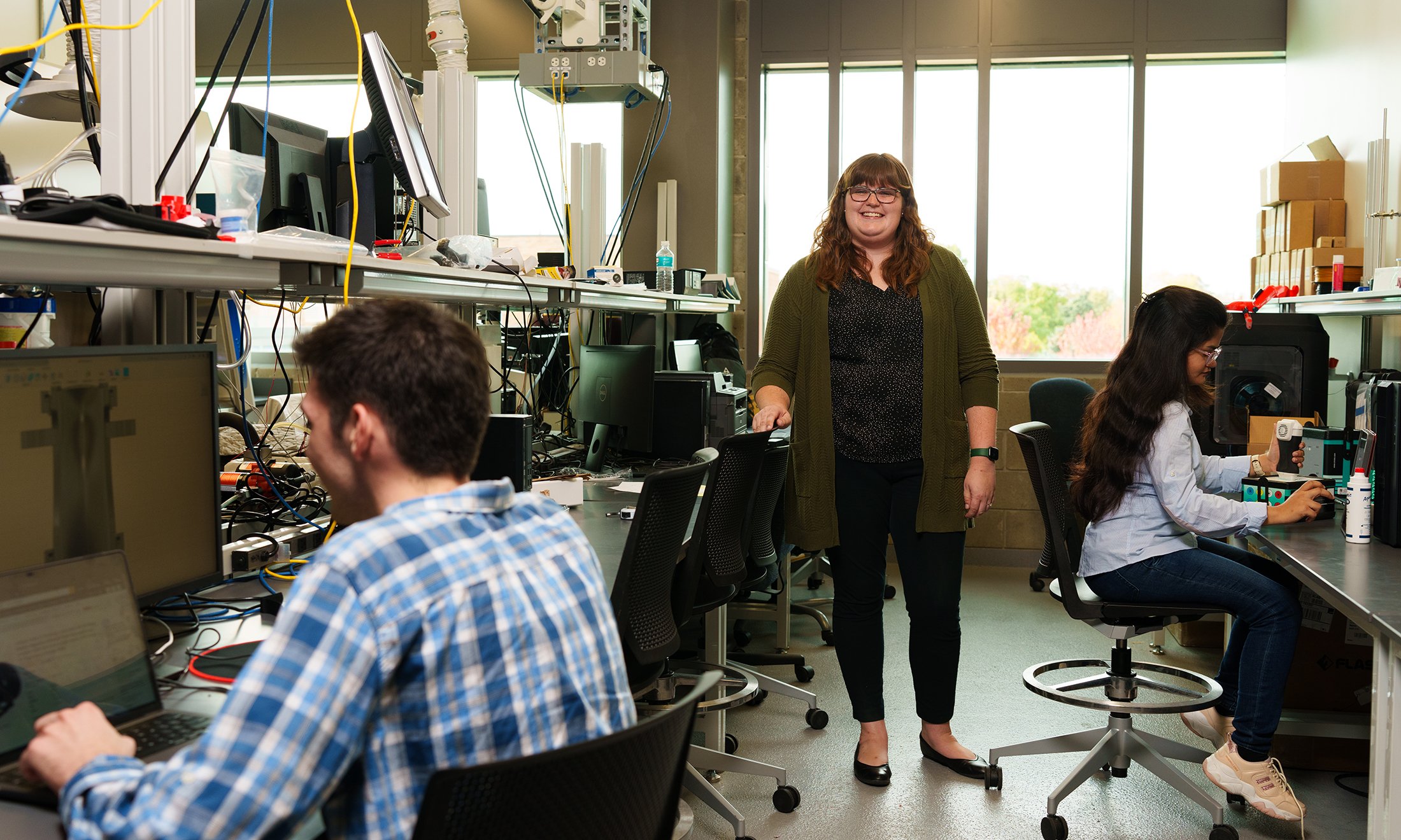
Rehnuma Hasnat (M.S.) and undergraduate bioengineering students Luc Taburet and Molly Vue (not pictured) work with Dr. Wiacek on the project that pairs artificial intelligence with ultrasound and elastography to improve the diagnosis and screening for breast cancer.
Department of Bioengineering
 December 20, 2023
 By Arina Bokas
“Through my research, I strive to improve ultrasound technologies to create a new paradigm of acoustic-based screening methods for various diseases,” says Alycen Wiacek, Ph.D., OU assistant professor, who works across two departments — the Department of Bioengineering and the Department of Electrical and Computer Engineering. “When combined with the correct hardware for signal detection and software for image combination and display, acoustic-based technologies can pave the way to a new generation of painless, ionizing radiation-free, and low-cost screening methods,” she adds.
Already as a Ph.D. student at Johns Hopkins University, Dr. Wiacek made considerable gains towards improving diagnostic quality and patient care by leveraging AI and other advanced signal processing techniques to develop systems in ultrasound and photoacoustic imaging. She implemented a novel deep learning architecture, which learned physics-based features directly from raw ultrasound data, and created a novel photoacoustic imaging-based surgical guidance system for safer hysterectomy procedures.
In her second year at Oakland University, Dr. Wiacek’s laboratory, the Medical Acoustics for Global health Imaging and Clinical translation (MAGIC) lab, continues strengthening SECS expertise in biomedical imaging and AI.
One of her current projects, supported by a grant from NASA through the Michigan Space Grant Consortium, focuses on the design of a digital optical and acoustic model of thrombosis. More colloquially known as a blood clot, this model can be used to determine the optimal combination of quantitative image-based features to characterize blood clots over time.
“In 2019, a clot was found blocking the internal jugular vein of an astronaut while in space, prompting investigations into the impact of microgravity on thrombosis. In settings like the international space station, there are limited resources to make such a critical diagnosis. Ultrasound is the ideal imaging method for such detection, but standard brightness mode (B-mode) ultrasound alone cannot quantitatively track blood clots over time or characterize them,” Dr. Wiacek explains.
Quantitative ultrasound, on the other hand, can leverage the acoustic scattering properties of tissue to understand and characterize aspects of tissue microstructure. Coupled with photoacoustic imaging, which leverages optical absorption properties of tissue, it has the potential to non-invasively provide quantitative information about the state and composition of the blood clot. Long term, these quantitative methods could be implemented in ex vivo and eventually in vivo settings, enabling monitoring of thrombosis over time in resource-limited settings in space and on Earth.
Another research area that Dr. Wiacek is currently investigating pairs artificial intelligence with ultrasound and elastography to improve the diagnosis and screening for breast cancer.
Elastography, which relies on the measurement of tissue motion before and after a small applied force, is a promising method to complement standard B-mode ultrasound. It enables the assessment of the stiffness of masses embedded in tissue. However, both the B-mode and elastography images still suffer from noise and artifacts caused by wave propagation through tissue layers.
“By adding specific types of noise to simulated ultrasound and elastography images we can simulate real clinical imaging scenarios. We then train the network by giving it both the noisy image and the clean image, allowing it to learn what the noise looks like and how to remove it. Consequently, when the network is given a new image, it is able to effectively remove the noise and improve the overall image quality.” says Dr. Wiacek.
“The neural network we are designing leverages information from both the traditional B-mode ultrasound image and the elastogram to improve image quality in both modalities and ultimately better visualize breast masses,” says Molly Vue, bioengineering undergraduate student, who works with Dr. Wiacek on the project and has recently won the Undergraduate Poster Presentation Award in Engineering at the 2023 Sigma Xi International Forum on Research Excellence.
The goals of the study are to create a dataset of simulated and phantom ultrasound and elastography images and develop an optimized network architecture to improve image quality. At the conclusion of the study, Dr. Wiacek plans to release all code and datasets publicly, enabling future benchmark evaluations. This study is funded by the OU University Research Committee.
“With proper engineering design, acoustic-based technologies are low cost, painless, and portable, making them the future of many disease screening solutions. Dr. Wiacek’s work is enabling this future. Through creating acoustic-based screening hardware using multimodal integration techniques, optimizing this hardware for seamless transition into the clinic, developing and optimizing algorithms for better image display, and building AI models, together, she is sculpting the future of equitable solutions,” states Shailesh Lal, Ph.D., Department of Bioengineering professor and chair.
Dr. Wiacek’s research is closely tied to the missions of several groups within the National Science Foundation and the National Institute of Health. Learn more information about her research and current projects.